Packaging of DNA into Chromatin and Chromosomes
Chromosomes are long double stranded DNA molecules. Over 150,000,000 nucleotide pairs make up the human X chromosome. The complete replication and orderly transfer of something this large requires the chromosome to be packaged for stability and organization which creates genetic stability in the cells of a species. Every cell in a diploid organism contains two copies (2n) of every chromosome present in that organism (Fig. 5). For example, humans have 46 chromosomes in their body, 23 were inherited from the father and 23 from the mother. Gametes, the reproductive cells of an organism (egg or sperm), have only one set (1n) of chromosomes. When the two gametes unite, they form a living embryo with two sets of genetic information. The packaging of DNA into a stable chromosome is essential to ensure that organisms retain all of their inherited genetic information in all of their cells.
Figure 5. Sets of chromosomes in cells. Every cell contains chromosomes. Chromosomes can be thought of as sets of cards. Haploid cells (1n) contain one set of cards, meaning one set of chromosomes. Diploid cells (2n) contain two sets of cards, or two sets of chromosomes. Diploid cells (2n) contain two sets of cards, or two sets of chromosomes. Credit: NIH-NHGRI.
Inside cell nuclei, DNA is assembled, or packaged, into material called chromatin, which is composed of both DNA and proteins (Figure 6). Chromatin functions to protect and regulate DNA, as well as to efficiently store the very long DNA molecules that make up chromosomes within the limited space of the nucleus.
DNA from higher eukaryotic organisms is about 109 to 1010 base pairs (bp). For example, human and maize chromosomal DNA is about 109 bp, which is equivalent to 1.8 meters (m) if all chromosomal DNA were stretched end to end in a linear manner. The diameter of the nucleus is only about 4-6 μm. All of the DNA in the genome fits into the organism’s nucleus because it is packaged and condensed into chromatin fibers which is DNA binding with specialized structural proteins called histones (Fig. 6). Chromatin fibers can be observed in the nucleus of cells with an electron microscope (10,000,000X power). Packing into a condensed structure is even more critical during cell division. The chromosome must be condensed into structures that are small and mobile. These are the chromosomes that are observed under a light microscope (100X power, Figure 7).
Figure 6. Chromosome structure. DNA is packed into chromatin fibers and chromosomes. Chromatin fibers are histones bound in double helix DNA. Chromatin by NIH-NHGRI, n.d. Public Domain.
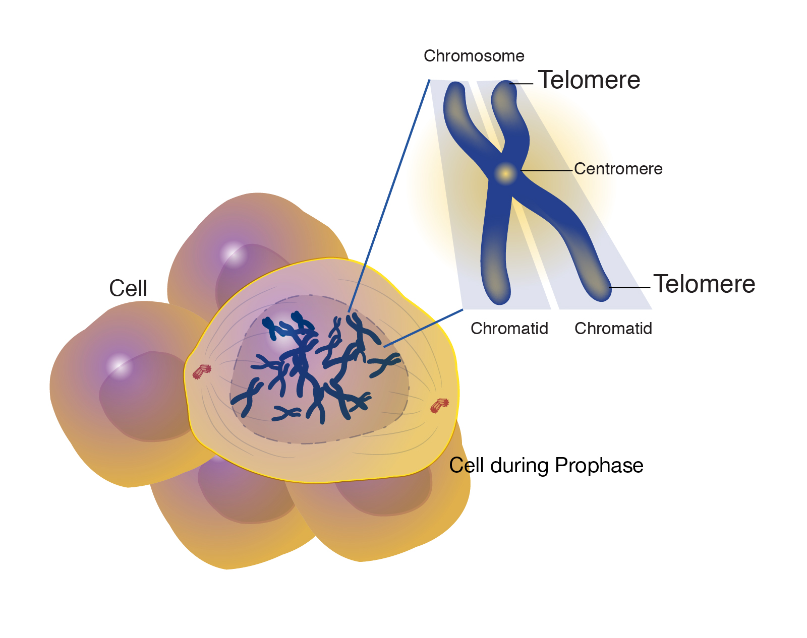
Figure 7. Chromosome structures during prophase. Chromosomes have centromeres toward the middle of a chromosome and telomeres at each end of the chromosome. Telomere by NIH-NHGRI, n.d. Public Domain.
Centromeres
The centromere is a chromosomal region that controls chromosome segregation at mitosis and meiosis (Fig. 7). Centromeres connect to microtubules of the spindle apparatus, which directs their movement to opposite poles (daughter cell nuclei) during cell division. Scientists were successful in isolating these centromeric sequences from yeast and engineered brewer’s yeast (S. cerevisiae) plasmids that were able to replicate like the chromosomes. Through these efforts scientists were able to point centromeric function to a DNA stretch of about 120 bp that was resistant to DNase (deoxyribonuclease) and bound to a single microtubule.
Telomeres
The telomere lies at the end of the chromosome and confers stability by “sealing” the end of a chromosome (Fig. 7). Telomeres consist of a long series of short, repeated DNA sequences that occur in tandem arrays and are added to the end of the chromosome during DNA replication by an enzyme called telomerase. In most plant species the sequence TTTAGGG constitutes a conserved telomere motif.